
Pin by Deborah on Painting Dog portraits painting, Animal paintings, Animal drawings
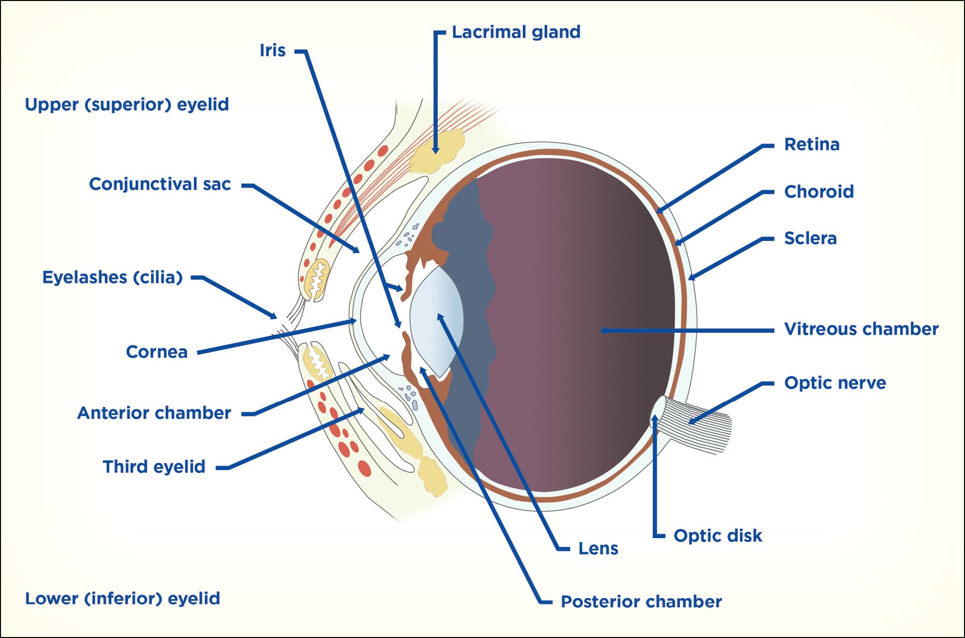
When considering the anatomy of the eye, it is important to first consider the orbit as a whole.. In the dog, the upper eyelid contains >2 rows of cilia, whereas the cat does not have any. Instead the first row of hairs on the skin are adapted for the same purpose. There are two types of gland associated with the cilia; Moll (which are.
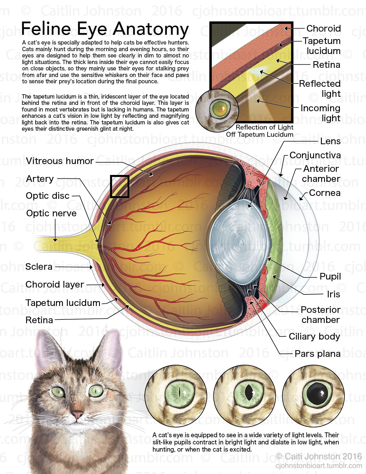
Scientific Illustration cjohnstonbioart Feline Eye Anatomy (2016)
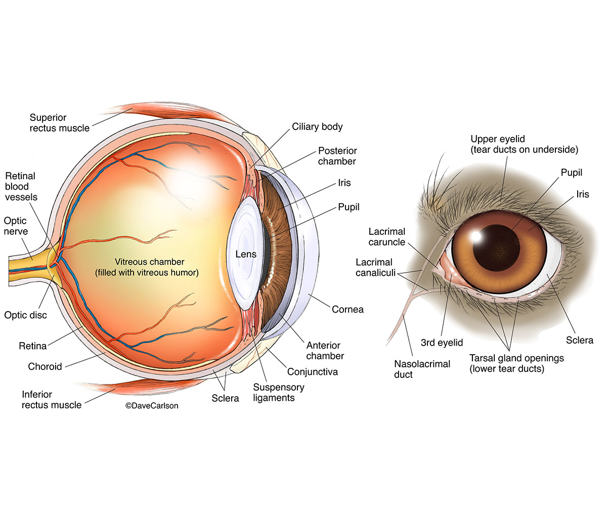
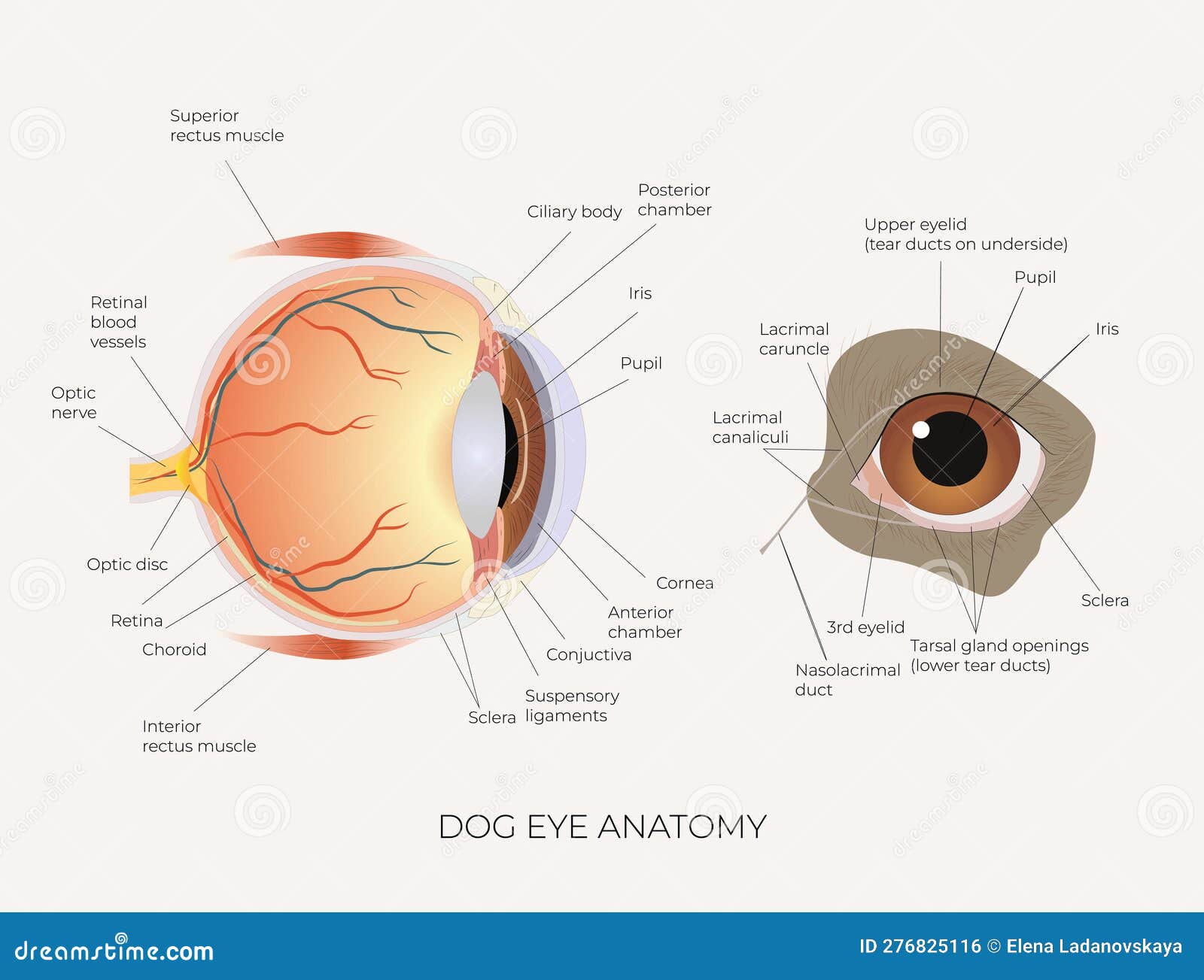
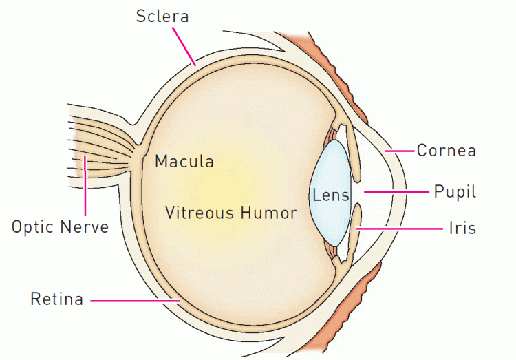
The anatomy of a dog's eye is very similar to that of a human's, though vision differs greatly. Cornea: A thin, smooth, transparent layer at the front of the eye. Trauma, ulceration, and irritating chemicals can lead to changes in the clarity of the cornea. Sclera: The "whites of the eyes."

lacrimal caruncle Google 검색 Eye anatomy, Dog eyes, Cherry eye in dogs
The anatomy of dogs' and cat's eyes work by adjusting to different light conditions essential for hunting or tracking prey. Important Structures of the Eye The orbit is the bony cavity or socket which holds the eyeball as well as muscles, nerves, blood vessels, and structures that produce and drain tears.

Dog eye anatomy explained Brookfield Animal Hospital
Let's take a look at the anatomy of a dogs eye and how a dog's eyesight compares to ours—from seeing colors to side vision and seeing in the dark. Dog Eye Anatomy. The anatomy of a dog's eye is very similar to that of a human eye. Dogs have an upper and lower eyelid, the same as people. There are many other similarities, including:
Canine Uveitis and the Veterinary Technician Today's Veterinary Nurse
General Anatomy. Dogs generally have the same anatomical structure as humans [2] Dogs have a third eyelid, called the nictitating membrane, which further protects the eye, spreads tearfilm over the cornea, and is involved in tear production. [2] Dogs have a tapetum lucidum behind the retina, which is highly reflective, and increases night vision.
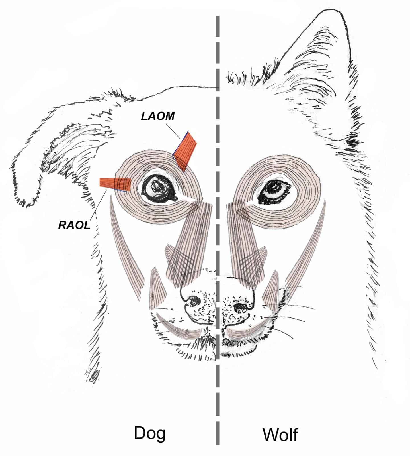
How a unique facial muscle makes those 'puppy dog eyes' irresistible to humans
The eye ( organum visus) (Fig. 21-1) develops as a neuroectodermal outgrowth of the embryonic prosencephalon that contacts surface ectoderm and is enveloped by induced mesodermal and neural crest mesenchyme. The definitive eye and its adnexa are contained within an orbit that is only partly bony.
.png)
Dry Eye/KCS Little Critters Veterinary Hospital Gilbert, AZ
Dog Eye Anatomy " Dog Eye Anatomy is a reflection of the eye as a complicated organ, containing many parts. These functional parts, i.e. sclera, conjunctiva, cornea, iris, pupil, lens, retina, lacrimal gland etc. are contained in a bony socket called a "orbit".
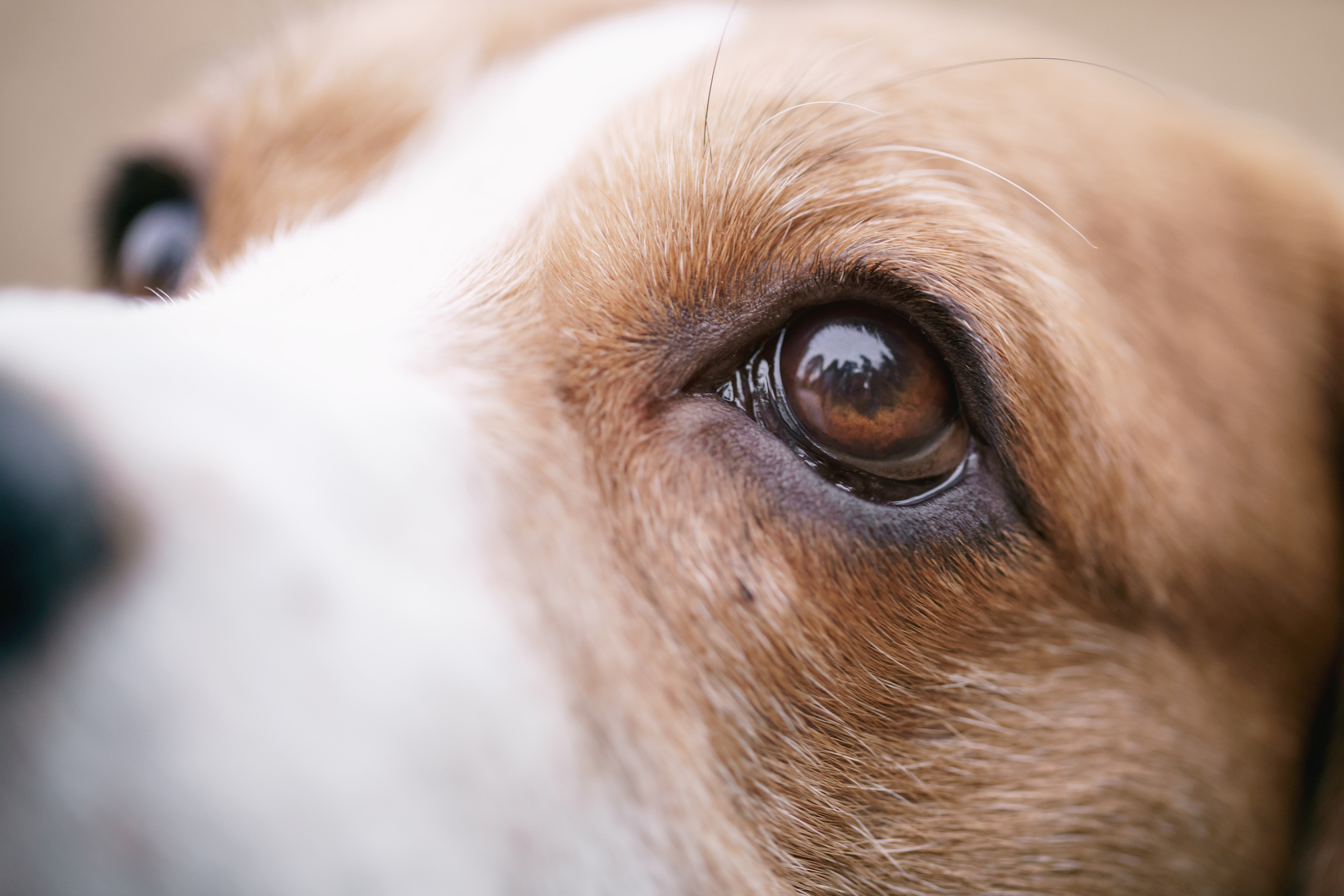
Dog Eye Photograph by Laurie OKeefe
Pet Eye Disease EYE ANATOMY AND DISEASE MASTER CLASS™ Introduction Vision is one of the five senses along with taste, smell, hearing, and touch. The eyes are direct extensions of the brain, receiving visual information and processing it to give a stereoscopic view of the world.
Dog Eye Anatomy Carlson Stock Art
Dog eye anatomy Dogs don't have a mere two eyelids—they have three. While your dog may blink with two eyelids, just like we do, they also have a third lid, called a nictitating membrane or a haw, which moves horizontally across the eye from the inside corner.
Dog Eye Anatomy Science Education Poster Stock Vector Illustration of medicine, zoology 276825116
To understand the anatomy of the canine eye in "A Closer Look at the Canine Eye: Understanding Your Dog's Vision", you need to explore the structure of the eye, understand the function of different parts of the eye, and compare it to the human eye.
Dog eye anatomy Quiz
A T-shaped cartilage that forms a firm connective tissue A tear gland held in place by the T-shaped cartilage A dog's third eyelid extends across the eye to: Protect the eyes from scratches, especially if the dog is walking or running through brush or somewhere the eye may get scratched Keep the eyes moist by spreading tears across the eyeball.

Why are my dog's eyes goopy? — VAH
The dog eye anatomy includes eyeball, orbit, eyelid, and lacrimal apparatus. This article will help you to know the details of these structures from the dog eye with labeled diagrams. Quick overview: The eyeball of a dog eye is located in the orbital cavity and consists of 3 tunics - fibrous, vascular, and nervous.
Dog Eye Colors Coats and Colors
1 KEEP YOUR DOG'S EYES HEALTHY: OVERVIEW 1. Eyes respond well to natural health prevention methods, so keep them healthy with nutrition, exercise, care of the immune system, and avoidance of toxins and stressors. 2. Use alternative therapies - by themselves or in combination with conventional medicine - to treat short- or long-term eye problems.

Dogs Eye Anatomy Everything You Need To Know About Them Zigzag
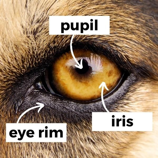
Dog Vision: Understanding Canine Eye Anatomy Dogs' eyes are structurally very similar to human eyes. The colored part is the iris, which surrounds the dark round pupil and controls how much light passes through that opening.
Dog Eye Anatomy Third Eyelid dogjullle
Dog eyes are made up of a cornea, iris, pupil, lens, retina, and sclera. They also have an upper and lower eyelid and a third eyelid on the outside of the eye for protection. Rods and cones are how images and light are processed and important for vision. Let's take a closer look at how each of these works. Cornea
All About Your Dog's Eyes
What Is the General Structure of the Canine Eye? The eye is formed by three concentric (circular) tunics or layers of tissue. These are the outer fibrous tunic, the middle vascular tunic and the inner nervous tunic.